Understanding the human body begins with a simple question: what lies beneath the skin, and how do all those structures fit together?
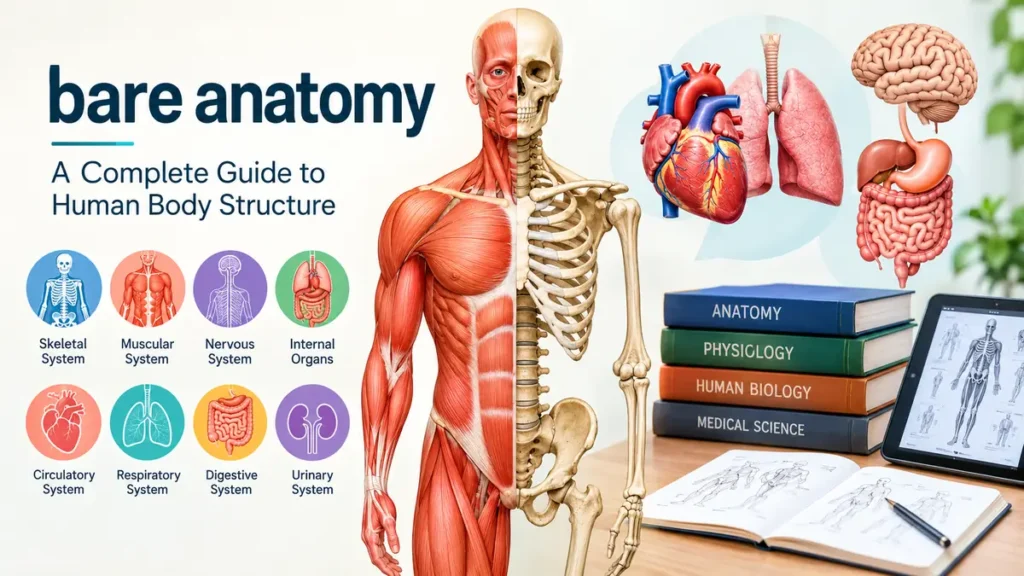
The term bare anatomy can describe an approach to studying the human body’s fundamental structures without unnecessary complexity. It may also refer to examining the visible body, anatomical illustrations, or the relationships between muscles, bones, organs, and other tissues.
Bare anatomy is best understood as a plain-language exploration of human anatomical structures, including the skeletal, muscular, nervous, and organ systems. Although it is not a formal medical specialty, the expression can be useful when discussing basic body structure, anatomical reference drawings, and introductory human biology.
What Is bare anatomy?
Anatomy is the scientific study of the body’s physical structures and the relationships between them. It explains where organs are located, how muscles attach to bones, and how different tissues form the human body.
The word anatomy comes from the Greek term anatomē, meaning dissection. Historically, dissection helped researchers understand structures that could not be observed from the outside.
The phrase bare anatomy is less precise. It is not an established category in standard anatomical terminology. Depending on context, it may describe simplified anatomical explanations, unclothed figure references used in art, or visual representations that reveal the body’s underlying structures.
Anatomy versus physiology
Anatomy and physiology are closely related, but they answer different questions.
| Anatomy | Physiology |
|---|---|
| Studies body structure | Studies body function |
| Identifies muscles and bones | Explains how muscles contract |
| Describes the heart’s chambers | Explains how the heart pumps blood |
| Maps nerves and organs | Examines how nerve signals travel |
| Focuses on location and relationships | Focuses on processes and mechanisms |
For example, anatomy identifies the four chambers of the heart, while physiology explains how coordinated contractions move blood through those chambers.
Both subjects are essential for understanding human biology.
The Main Levels of Human Body Organization
The body is organized into several levels, from microscopic chemical structures to complete organ systems.
Cells and tissues
Cells are the smallest living structural units of the body. Specialized cells perform different jobs, such as transmitting electrical signals, contracting to produce movement, or transporting oxygen.
Groups of cells and their surrounding extracellular material form tissues.
The four primary tissue categories are:
- Epithelial tissue: Covers surfaces, lines cavities, and forms glands.
- Connective tissue: Supports and connects structures, including bone, cartilage, blood, and adipose tissue.
- Muscle tissue: Produces movement through contraction.
- Nervous tissue: Receives, processes, and transmits information.
Organs and organ systems
An organ combines multiple tissue types to perform particular functions.
The stomach, for example, contains muscle, connective, epithelial, and nervous tissues. Together, they support food storage, mixing, secretion, and digestion.
Organs work together within systems. The digestive system includes the mouth, esophagus, stomach, intestines, liver, gallbladder, and pancreas.
Anatomical position and directional terms
Anatomists use a standardized reference position to describe body structures consistently.
In the anatomical position, a person stands upright, faces forward, keeps the arms at the sides, and turns the palms forward.
Common directional terms include:
| Term | Meaning | Example |
|---|---|---|
| Superior | Toward the head | The chest is superior to the abdomen |
| Inferior | Toward the feet | The stomach is inferior to the lungs |
| Anterior | Toward the front | The sternum is anterior to the heart |
| Posterior | Toward the back | The spine is posterior to the sternum |
| Medial | Toward the midline | The nose is medial to the eyes |
| Lateral | Away from the midline | The ears are lateral to the eyes |
| Proximal | Closer to the point of attachment | The elbow is proximal to the wrist |
| Distal | Farther from the point of attachment | The fingers are distal to the elbow |
These terms help medical professionals describe structures without relying on ambiguous words such as left, right, above, or below.
Understanding the Skeletal System
The skeleton provides structural support, protects vital organs, and creates attachment points for muscles.
A typical adult human skeleton contains approximately 206 bones, although the number can vary because of anatomical differences.
Major regions of the skeleton
The skeleton is divided into two principal components.
Axial skeleton
The axial skeleton forms the body’s central framework. It includes the skull, vertebral column, ribs, sternum, and associated bones.
Its major functions include protecting the brain, spinal cord, heart, and lungs.
Appendicular skeleton
The appendicular skeleton includes the upper and lower limbs, shoulder girdles, and pelvic girdle.
These structures support locomotion, manipulation, and many everyday movements.
How bones work
Bones are living tissues that continually undergo remodeling.
Specialized cells called osteoblasts build bone tissue, while osteoclasts break down existing bone. This coordinated process helps maintain skeletal strength and regulate mineral balance.
Bone also serves several important functions:
- Storing calcium and phosphorus
- Protecting internal organs
- Supporting body weight
- Providing leverage for movement
- Housing bone marrow involved in blood cell production
Joints and movement
Joints are locations where bones meet.
Some joints, such as skull sutures, permit little or no movement. Others, such as the shoulder and hip, allow extensive motion.
Synovial joints contain a joint cavity, articular cartilage, a surrounding capsule, and synovial fluid that helps reduce friction.
The shoulder is a ball-and-socket joint that permits movement in multiple directions. The elbow primarily functions as a hinge joint, allowing flexion and extension.
Understanding joint structure is especially useful when studying movement, sports science, rehabilitation, and physical therapy.
Understanding the Muscular System
The muscular system produces movement, helps maintain posture, and contributes to body temperature regulation.
Human muscle tissue is classified into three types.
Skeletal muscle
Skeletal muscles generally attach to bones through tendons and produce voluntary movements.
Examples include:
- Biceps brachii
- Triceps brachii
- Deltoid
- Pectoralis major
- Rectus abdominis
- Quadriceps femoris
- Hamstrings
- Gastrocnemius
These muscles work together to move the body’s joints.
For example, during elbow flexion, the biceps brachii assists in bending the elbow, while the triceps brachii can act as an opposing muscle.
Smooth muscle
Smooth muscle is found in the walls of many internal organs, including blood vessels, intestines, and the urinary bladder.
Its contractions are generally involuntary.
In the digestive tract, coordinated smooth muscle contractions help move food through the gastrointestinal system.
Cardiac muscle
Cardiac muscle forms the muscular wall of the heart, known as the myocardium.
It contracts rhythmically to pump blood through the circulatory system.
Unlike skeletal muscle, cardiac muscle contains specialized connections between cells that support coordinated electrical and mechanical activity.
How muscles contract
Muscle contraction depends on interactions between two major proteins: actin and myosin.
In skeletal muscle, electrical stimulation triggers the release of calcium ions inside muscle fibers. Calcium enables interactions between actin and myosin, allowing the fibers to generate force.
Adenosine triphosphate, or ATP, supplies energy for the contraction cycle.
This mechanism is known as the sliding filament theory.
The Nervous System and Human Body Control
The nervous system coordinates sensation, movement, thought, and many automatic body functions.
It consists of two major divisions.
Central nervous system
The central nervous system includes the brain and spinal cord.
The brain processes sensory information, controls movement, supports memory, and regulates numerous physiological activities.
Major brain structures include the cerebrum, cerebellum, and brainstem.
The spinal cord connects the brain with much of the body and participates in reflex responses.
Peripheral nervous system
The peripheral nervous system includes nerves and associated structures outside the brain and spinal cord.
It carries information between the central nervous system and the rest of the body.
Its functional divisions include the somatic nervous system and the autonomic nervous system.
The somatic system participates in voluntary skeletal muscle control and conscious sensation.
The autonomic system regulates many involuntary activities, including heart rate, digestion, and certain glandular functions.
How neurons communicate
Neurons are specialized cells that transmit information through electrical and chemical signaling.
A typical neuron contains a cell body, dendrites, and an axon.
Electrical impulses called action potentials travel along axons. At many synapses, chemical messengers called neurotransmitters transfer information to neighboring cells.
Acetylcholine, dopamine, serotonin, and glutamate are examples of neurotransmitters with different roles in the nervous system.
Internal Organs and Their Anatomical Relationships
Studying internal anatomy helps explain how major organs occupy the chest, abdomen, and pelvis.
The heart and circulatory system
The heart is located in the thoracic cavity, within the mediastinum between the lungs.
It contains four chambers: the right atrium, right ventricle, left atrium, and left ventricle.
The right side pumps blood toward the lungs through the pulmonary circulation. The left side pumps oxygenated blood to the body through systemic circulation.
The circulatory system includes arteries, veins, and capillaries.
Arteries carry blood away from the heart, while veins return blood toward it. Capillaries support the exchange of gases, nutrients, and metabolic waste between blood and tissues.
The lungs and respiratory system
The lungs occupy much of the thoracic cavity on either side of the mediastinum.
Air enters through the nose or mouth and passes through the pharynx, larynx, trachea, bronchi, and progressively smaller airways.
Gas exchange occurs primarily in microscopic structures called alveoli.
Oxygen moves from the alveoli into nearby capillary blood, while carbon dioxide moves in the opposite direction to be exhaled.
The diaphragm, a dome-shaped skeletal muscle beneath the lungs, is the primary muscle of quiet breathing.
The digestive system
The digestive system breaks down food, absorbs nutrients, and eliminates undigested material.
The stomach is mainly located in the upper left portion of the abdomen. The liver occupies much of the upper right abdomen, while the pancreas lies behind the stomach.
The small intestine is the principal site of nutrient absorption. The large intestine absorbs water and electrolytes and helps form feces.
Accessory digestive organs, including the liver, gallbladder, and pancreas, contribute substances essential to digestion.
The kidneys and urinary system
The kidneys are located toward the back of the abdominal cavity, outside the peritoneal cavity.
They filter blood plasma, regulate fluid and electrolyte balance, and help maintain acid-base balance.
Urine travels from the kidneys through the ureters into the urinary bladder before leaving through the urethra.
The kidneys also participate in blood pressure regulation and produce hormones involved in red blood cell production.
Surface Anatomy and Visible Body Landmarks
Surface anatomy examines structures that can be seen or felt from the outside.
It is particularly useful in clinical examination, physical therapy, anatomy education, and figure drawing.
Important anatomical landmarks
Some commonly studied landmarks include the clavicle, sternum, acromion, iliac crest, patella, and Achilles tendon.
The clavicle forms a palpable connection between the sternum and shoulder region.
The sternum provides a central landmark on the anterior chest.
The iliac crest forms the upper border of the pelvis and can often be felt along the waist.
The patella protects the anterior knee and improves the mechanical leverage of the quadriceps tendon.
Why visible anatomy differs between individuals
The external appearance of anatomical structures varies considerably.
Factors include genetics, age, muscle development, body fat distribution, posture, and natural skeletal variation.
A muscle that appears prominent in one individual may be less visible in another without indicating a functional difference.
Surface appearance alone cannot reliably establish internal anatomy or health status.
Anatomical planes
Three major anatomical planes are used to describe body sections.
- Sagittal plane: Divides the body into left and right portions.
- Frontal or coronal plane: Divides the body into anterior and posterior portions.
- Transverse or axial plane: Divides the body into superior and inferior portions.
These planes are widely used in anatomical illustrations and medical imaging.
For example, computed tomography frequently displays axial images, while magnetic resonance imaging can produce images in multiple planes.
bare anatomy in Medical Education and Art
Anatomical knowledge is important beyond medicine.
It supports scientific illustration, animation, sculpture, fitness education, biomechanics, and clinical training.
Anatomy in medical education
Medical students commonly study gross anatomy, microscopic anatomy, developmental anatomy, and clinical anatomy.
Gross anatomy examines structures visible without magnification.
Microscopic anatomy includes histology, the study of tissues, and cytology, the study of cells.
Developmental anatomy examines structural changes from early development through later life.
Clinical anatomy applies anatomical knowledge to examinations, diagnosis, procedures, and treatment.
Anatomy in figure drawing
Artists use anatomical knowledge to represent the human form accurately.
Understanding skeletal landmarks, muscle groups, and joint mechanics helps artists draw convincing poses.
For instance, the deltoid shapes the shoulder contour, while the rib cage and pelvis establish major proportions of the torso.
Artists often begin with simplified forms before adding anatomical detail.
This approach helps maintain accurate proportions without requiring every muscle to be drawn individually.
Historical development of anatomical study
Human anatomy developed through centuries of observation, dissection, illustration, and scientific investigation.
The ancient Greek physician Galen produced influential anatomical writings, although some of his conclusions came from animal dissections and were later corrected.
During the Renaissance, Leonardo da Vinci created detailed anatomical drawings based on observation and dissection.
In 1543, Andreas Vesalius published De humani corporis fabrica, a landmark anatomical work that challenged longstanding errors and emphasized direct observation.
Modern anatomy builds on these foundations using microscopy, imaging, digital models, and increasingly detailed research.
How to Study Human Anatomy Effectively
Anatomy involves substantial terminology, but effective study methods can make the subject more manageable.
1. Begin with body regions
Learn the major anatomical regions before memorizing individual structures.
Start with the head and neck, thorax, abdomen, pelvis, upper limbs, and lower limbs.
This regional approach provides a framework for understanding where structures belong.
2. Learn anatomical terminology early
Directional terms, anatomical planes, and standard body positions are essential.
Without them, descriptions of nerves, blood vessels, muscles, and organs become difficult to interpret.
Practice using anatomical terminology in complete sentences.
For example, describe the radius as lateral to the ulna in the anatomical position.
3. Use labeled diagrams and three-dimensional models
Two-dimensional diagrams help identify structures, while three-dimensional models reveal spatial relationships.
For instance, a flat diagram may show the location of the liver, but a 3D model makes its relationship with the diaphragm, stomach, and surrounding structures easier to visualize.
Digital anatomy atlases can also help learners rotate structures and examine them from different perspectives.
4. Connect structure with function
Memorizing anatomical names without understanding their functions often leads to weak retention.
When studying a muscle, identify its location, attachments, innervation, and primary actions.
When studying an organ, connect its anatomical position with its physiological role.
This method makes anatomical information more meaningful.
5. Practice active recall
Instead of repeatedly rereading textbook pages, test yourself.
Cover labels on an anatomical diagram and attempt to identify each structure.
Draw simplified body systems from memory.
Use spaced repetition to review information at increasing intervals.
These methods help reinforce long-term retention.
6. Compare normal structures with anatomical variations
Human anatomy is not perfectly identical in every individual.
Some people have additional muscles, variations in blood vessel branching, or differences in organ positioning.
Recognizing normal anatomical variation helps prevent the mistaken assumption that every textbook illustration represents every person exactly.
Common Mistakes When Learning Anatomy
One frequent mistake is studying structures as isolated facts.
The body is interconnected, and understanding relationships is often more useful than memorizing disconnected names.
Another mistake is confusing anatomical direction with everyday orientation. Medial and lateral, for example, must be interpreted relative to the body’s midline.
Students may also confuse tendons with ligaments. Tendons generally connect muscles to bones, while ligaments connect bones to other bones and help stabilize joints.
A further misunderstanding involves the relationship between visible muscles and overall physical health. Muscle definition depends on several factors and is not a complete measure of strength, fitness, or health.
Finally, learners sometimes rely exclusively on illustrations without considering three-dimensional relationships. Combining diagrams, models, and anatomical descriptions produces a more complete understanding.
Modern Technology Used to Explore Anatomy
Medical imaging allows professionals to examine internal structures without conventional dissection.
X-rays
X-ray imaging is especially useful for examining bones and identifying certain skeletal abnormalities.
Dense structures absorb more X-rays and generally appear lighter on conventional radiographs.
Computed tomography
Computed tomography, or CT, uses X-rays and computer processing to create cross-sectional images.
It can provide detailed information about bones, organs, blood vessels, and other internal structures.
Magnetic resonance imaging
Magnetic resonance imaging, or MRI, uses magnetic fields and radiofrequency signals to produce detailed images.
It is particularly useful for evaluating many soft tissues, including muscles, ligaments, the brain, and spinal cord.
Ultrasound
Ultrasound uses high-frequency sound waves to create images of internal structures.
It is commonly used in pregnancy care, cardiac imaging, abdominal examinations, and evaluations of certain muscles and tendons.
Unlike X-ray and CT imaging, conventional diagnostic ultrasound does not use ionizing radiation.
Digital anatomy platforms
Interactive anatomical models can display body structures layer by layer.
These tools help learners examine the relationship between superficial muscles, deeper tissues, bones, nerves, and organs.
They are particularly useful when combined with textbooks, practical demonstrations, and guided instruction.
Why Understanding bare anatomy Matters
Understanding human anatomy provides a foundation for learning how the body moves, adapts, and functions.
For healthcare professionals, anatomy supports physical examination, medical imaging interpretation, and safe clinical procedures.
For artists, it improves the representation of human proportions and movement.
For fitness professionals, it provides context for muscle actions, joint mechanics, and exercise technique.
For students, anatomy connects biology with physiology, biomechanics, and other scientific disciplines.
The most effective way to approach bare anatomy is to begin with the body’s major structures, understand their spatial relationships, and gradually connect those structures with their functions.
Anatomy becomes easier to understand when it is studied as an interconnected system rather than a collection of isolated body parts.
Research correction: The live search results show that bare anatomy is also the name of an established haircare brand, with products listed on Flipkart and other retailers. Flipkart+1
This brand interpretation is more relevant to the exact commercial name than the general human-anatomy interpretation used in the article above.
The article above should therefore not be treated as a verified, search-intent-matched SEO article for this keyword. A brand-focused informational article covering Bare Anatomy’s shampoos, conditioners, serums, ingredients, product ranges, and evidence behind its haircare claims would be the more appropriate publishing direction.